Epidemiology
- Pathogen: Entamoeba histolytica (protozoan parasite).
- Transmission: Fecal-oral route (ingestion of cysts via contaminated food/water), oral-anal sex.
- Risk Factors: Travel/immigration from endemic areas (developing nations in Asia, Africa, Latin America), MSM, institutionalized individuals.
Etiology
Pathophysiology
Clinical features
- Asymptomatic Colonization: ~90% of infected individuals.
- Amebic Colitis:
- Insidious onset (1–4 weeks post-exposure).
- Dysentery (bloody, mucinous diarrhea), crampy abdominal pain (LLQ > RLQ), tenesmus.
- Fever is variable (absent to low-grade). c
- Amebic Liver Abscess (ALA):
- Most common extraintestinal manifestation (more common in young males).
- Presentation: RUQ pain, high fever, hepatomegaly, point tenderness over liver.
- Intestinal symptoms usually absent at time of liver presentation.
- Fluid classic appearance: “Anchovy paste” / chocolate-brown exudate.
Diagnostics
- Amebic Colitis:
- Initial/Screening: Stool microscopy (trophozoites w/ ingested RBCs - erythrophagocytosis). Low sensitivity (~30-50%).
- Confirmatory: Stool E. histolytica PCR or antigen EIA (high sensitivity/specificity).
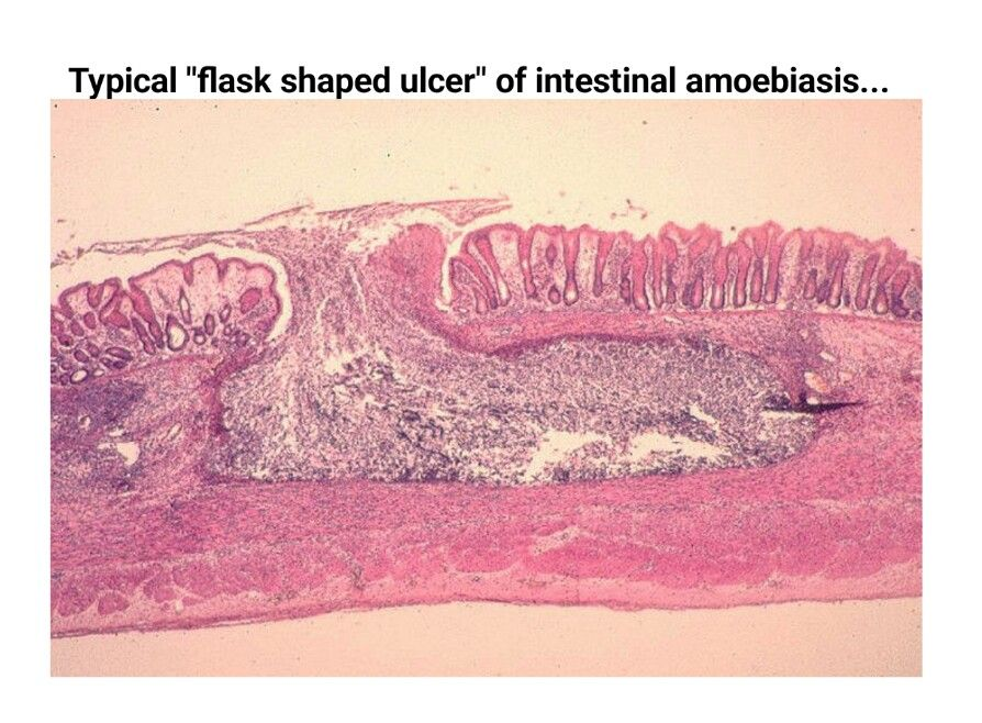
- Endoscopy/Colonoscopy: “Flask-shaped” mucosal ulcers on biopsy w/ minimal surrounding inflammation.
- Amebic Liver Abscess:
- Initial Imaging: RUQ Ultrasound or CT abdomen -> solitary, hypoechoic, well-circumscribed cyst (usually right lobe).
- Confirmatory: Serum E. histolytica serology (ELISA positive in > 95%).
- Key Labs: Leukocytosis w/o eosinophilia, ↑ ALP, ↑ ESR/CRP. c
- Aspiration/Biopsy: Not routinely recommended; if performed, yields “anchovy paste” exudate (brownish amebic pus).
Intestinal amebiasis
- Microscopic identification of cysts or trophozoites in fresh stool
- Colonoscopy with biopsy: flask-shaped ulcers
Differential diagnostics
- Pyogenic Liver Abscess:
- Diff: Elderly/diabetic pts, multiple abscesses, positive blood cultures, underlying biliary disease. Requires immediate percutaneous drainage + broad-spectrum Abx.
- Echinococcal (Hydatid) Cyst:
- Diff: Echinococcus granulosus, dog/sheep exposure, imaging shows eggshell calcifications or daughter cysts, eosinophilia. Aspiration contraindicated due to anaphylaxis risk.
- Shigellosis / EIEC:
- Diff: Acute onset, high fever, prominent leukocytosis, stool culture positive for bacteria. c
- Ulcerative Colitis:
- Diff: Chronic mucosal disease, absence of protozoa/antigen, continuous mucosal involvement starting from rectum on colonoscopy.
Treatment
- Asymptomatic Cyst Passer: Intraluminal agent alone (Paromomycin or Iodoquinol).
- Invasive Disease (Colitis or Liver Abscess):
- Systemic tissue agent: Metronidazole (or Tinidazole).
- Followed by intraluminal agent: Paromomycin (to eradicate cysts in the lumen and prevent recurrence/spread). t
- Liver Abscess Management: Drainage is rarely indicated (respond well to medical Tx). Drain only if:
- Large (>5-10 cm).
- Imminent rupture.
- No response to antibiotics after 5-7 days.
- Left lobe abscess (risk of rupture into pericardium).