Microscopic anatomy
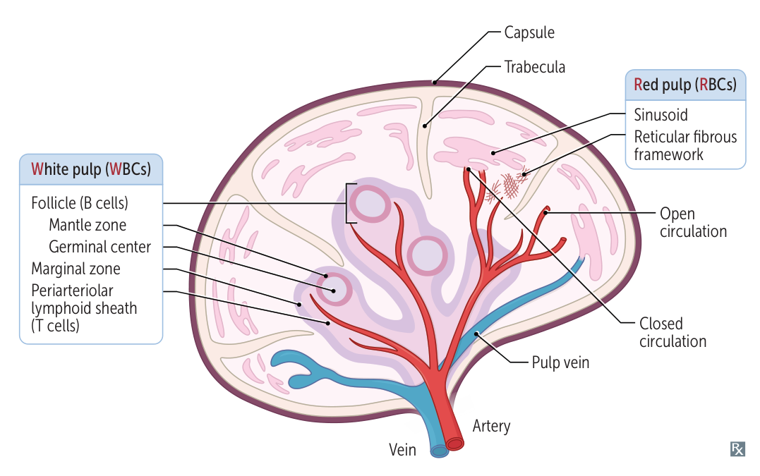
Red pulp
Red pulp is composed of splenic cords and sinuses (sinusoids).
- Splenic cords: a reticular meshwork filled with blood (open circulation system) that filters the blood from damaged erythrocytes
- Spleen sinusoids: long vessels with a fenestrated ring-like (“barrel hoop”) basement membrane that prevent old or malformed RBCs/platelets from reentering venous circulation
- Open circulation: Blood empties from sheathed capillaries into the splenic cords and then enters the sinusoids through slits in the vessel wall.
- Closed circulation: Blood empties from sheathed capillaries of the red pulp directly into the sinusoids.
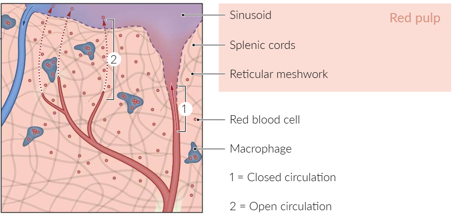
- Macrophages: found in the cords and around the sinusoids
- Phagocytosis of damaged RBCs/platelets that do not reenter circulation
- Capture of viruses and opsonized pathogens that enter the red pulp
- Blood flow: splenic artery → arterioles → red pulp (cords → sinusoids) → venules → splenic vein → portal circulation
White pulp
- Periarteriolar lymphatic sheath
- Surrounds the arterioles
- Dense lymphoid tissue containing T lymphocytes
- Splenic follicles
- Main component of white pulp
- Close to periarteriolar lymphatic sheath
- Contain B lymphocytes
- Splenic marginal zone
- Located between the red pulp and white pulp
- Contains antigen-presenting cells (APCs): macrophages and specialized B cells (marginal zone B cells)
Spleen Manifestations in Disease
The spleen’s primary roles are filtering blood and mounting immune responses. Its dysfunction or enlargement is a key sign of various systemic diseases. Spleen manifestations can be broadly categorized into Splenomegaly (enlargement) and Hyposplenism/Asplenia (decreased or absent function).
Splenomegaly (Enlarged Spleen)
- Etiologies of Splenomegaly
- Hematologic & Myeloproliferative Disorders
- Primary Myelofibrosis (PMF): Clonal megakaryocytes (JAK2), marrow fibrosis → massive EMH, dacrocytes (“teardrop” cells), dry tap.
- Hairy Cell Leukemia: Older males, BRAF V600E → massive splenomegaly + pancytopenia, dry tap, TRAP (+).
- Chronic Myeloid Leukemia (CML): BCR-ABL1 / t(9;22) → massive splenomegaly, basophilia, ↓ LAP score.
- Chronic Lymphocytic Leukemia (CLL): Elderly, asymptomatic absolute lymphocytosis, smudge cells, generalized LAD + HSM.
- Polycythemia Vera (PV): JAK2 mutation, hyperviscosity, aquagenic pruritus, facial plethora.
- Hodgkin & Non-Hodgkin Lymphoma: Direct tumor infiltration, “B symptoms” (fevers, night sweats, weight loss), painless LAD.
- Congestive & Vascular
- Cirrhosis / Portal HTN: Most common overall; red pulp pooling → congestive splenomegaly + secondary hypersplenism (thrombocytopenia).
- Splenic Vein Thrombosis: Post-pancreatitis or pancreatic CA → isolated gastric varices + splenomegaly (normal liver).
- Budd-Chiari Syndrome: Hepatic vein/IVC thrombosis → tender hepatomegaly, ascites, secondary splenomegaly.
- Hemolytic Anemias (RBC Clearance)
- Hereditary Spherocytosis (HS): Ankyrin/spectrin defect → spherocytes cleared in spleen; triad of anemia + jaundice + splenomegaly; (+) EMA test.
- Warm AIHA: IgG-mediated splenic RBC clearance; spherocytes, (+) Direct Coombs.
- Thalassemia Intermedia / Major: Severe ineffective erythropoiesis + chronic hemolysis → EMH, “crew-cut” skull X-ray, HSM.
- Infections
- EBV / CMV (Mononucleosis): Reactive lymphoid hyperplasia; high risk of splenic rupture (avoid sports ≥3–4 wks).
- Infective Endocarditis (IE): Immune clearance or septic emboli → splenic infarction/abscess (fever + LUQ pain).
- Visceral Leishmaniasis (Kala-azar): Sandfly (Leishmania) → massive splenomegaly + pancytopenia + spiking fevers.
- Malaria: Intravascular parasite cycling → hyperreactive malarial splenomegaly.
- Storage, Infiltrative & Autoimmune
- Gaucher Disease: β-glucocerebrosidase deficiency → massive splenomegaly, severe bone crises/AVN, “wrinkled tissue paper” macrophages.
- Niemann-Pick Disease: Sphingomyelinase deficiency → neurodegeneration, cherry-red macular spot + prominent HSM.
- Felty Syndrome: Long-standing Rheumatoid Arthritis (RA) + Neutropenia + Splenomegaly.
- Systemic Amyloidosis / Sarcoidosis: Splenic parenchyma infiltration by amyloid fibrils (Congo red +) or non-caseating granulomas.
- Hematologic & Myeloproliferative Disorders
- High-Yield “Massive Splenomegaly” (>8 cm / Crosses Midline)
- Primary Myelofibrosis
- Hairy Cell Leukemia
- Chronic Myeloid Leukemia (CML)
- Gaucher Disease
- Visceral Leishmaniasis (Kala-azar)
- Conditions WITHOUT Splenomegaly (High-Yield USMLE Traps)
- Immune Thrombocytopenia (ITP)
- Isolated severe thrombocytopenia (<30k) + mucosal bleeding.
- Autoantibody clearance occurs in spleen, but spleen is NOT enlarged (if splenomegaly is present, suspect CLL, SLE, or cirrhosis instead).
- Thrombotic Thrombocytopenic Purpura (TTP)
- Pentad: MAHA (schistocytes), Thrombocytopenia, Fever, AKI, Neurologic deficits.
- Microthrombi consume platelets in systemic microvasculature; normal spleen.
- Aplastic Anemia
- Severe pancytopenia + reticulocytopenia from marrow failure/autoimmune stem cell destruction.
- BM biopsy shows fatty hypocellular marrow; no EMH or splenomegaly (differentiates from PMF/leukemia).
- Sickle Cell Disease (Adults)
- Recurrent microvascular sickling and vaso-occlusion during childhood cause autosplenectomy (fibrotic, calcified nub by age ~6). c
- Spleen is ABSENT in adults (Howell-Jolly bodies on smear; contrast with young children who can have acute splenic sequestration crises).
- Multiple Myeloma (MM)
- CRAB criteria (Hypercalcemia, Renal failure, Anemia, Bone lytic lesions) + monoclonal M-spike (IgG/IgA).
- Plasma cell expansion is restricted to BM; NO splenomegaly or LAD (presence of HSM/LAD points to Waldenström Macroglobulinemia instead).
- Hemolytic Uremic Syndrome (HUS)
- MAHA + Thrombocytopenia + AKI following E. coli O157:H7 enteritis.
- Platelet consumption is localized to renal microthrombi; spleen is normal.
- Tay-Sachs Disease
- Hexosaminidase A deficiency → developmental regression, hyperreflexia, cherry-red macular spot.
- NO hepatosplenomegaly (the single most critical clinical differentiator from Niemann-Pick, which presents identically with HSM).
- Disseminated Intravascular Coagulation (DIC)
- Widespread microvascular thrombosis + consumptive coagulopathy (↑ PT, ↑ PTT, ↓ fibrinogen, ↑ D-dimer).
- Systemic intravascular consumption; spleen is normal.
- Immune Thrombocytopenia (ITP)
Hyposplenism & Asplenia
This refers to decreased or absent splenic function, leading to impaired blood filtering and immune response.
Pathophysiology/Etiology:
- Splenectomy: Surgical removal (e.g., for trauma, ITP, hereditary spherocytosis).
- Autosplenectomy: Repetitive infarction and fibrosis, classically seen in Sickle Cell Disease in adults.
- Congenital Asplenia: Rare developmental failure.
- Functional Hyposplenism: Spleen is present but dysfunctional (e.g., celiac disease, SLE, inflammatory bowel disease).
Diagnosis (Hallmark Peripheral Smear Findings):
- Howell-Jolly bodies: Basophilic nuclear remnants in RBCs (normally removed by spleen). Their presence is a key sign of hyposplenism.
- Target cells (Codocytes): Due to altered RBC membrane.
- Thrombocytosis: Loss of splenic sequestration of platelets.
- Acanthocytes (Spur cells): Spiky RBCs.
Complications & Management:
- Overwhelming Post-Splenectomy Infection (OPSI): High risk of fulminant sepsis from encapsulated organisms due to loss of opsonizing IgM production and phagocytosis. t
- Key Pathogens: Streptococcus pneumoniae (most common), Haemophilus influenzae type b, Neisseria meningitidis.
- Mnemonic: SHiN
- Prevention:
- Vaccinations: Administer vaccines against pneumococcus, meningococcus, and H. influenzae type b, ideally before elective splenectomy or as soon as possible after.
- Prophylactic Antibiotics: Often prescribed, especially for children and the immunocompromised (e.g., penicillin).
- Patient Education: Advise patients to seek immediate medical care for fevers.
| Disease | Spleen Finding | Mechanism |
|---|---|---|
| Sickle Cell Disease | Splenomegaly (in children) -> Autosplenectomy (in adults) | Vaso-occlusion from sickled RBCs causes initial splenic sequestration crisis, followed by recurrent infarctions leading to a shrunken, fibrotic spleen. |
| Hereditary Spherocytosis | Splenomegaly | ”Work hypertrophy” from clearing spherocytic RBCs. Splenectomy is curative. |
| Infectious Mononucleosis (EBV) | Splenomegaly | Lymphoid hyperplasia in response to viral infection. High risk of rupture. |
| Portal Hypertension | Congestive Splenomegaly | Increased pressure in the splenic vein obstructs outflow. |
| Myelofibrosis | Massive Splenomegaly | Extramedullary hematopoiesis (blood cell production shifts to spleen as bone marrow fails). |
| Hairy Cell Leukemia | Massive Splenomegaly | Infiltration of the red pulp by neoplastic B-cells. |
| ITP | Normal Spleen Size or Mild Splenomegaly | Spleen is the site of platelet destruction, but does not typically cause significant enlargement. |