Systemic: Fever, chills, tachycardia, altered mental status, leukocytosis (may be blunted in DM).
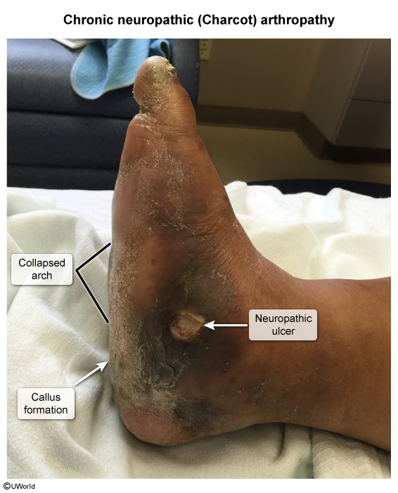
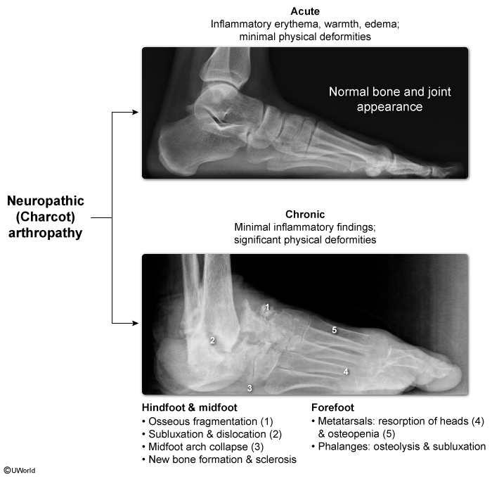
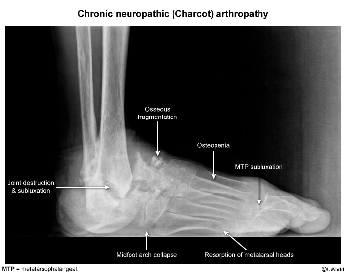
Charcot Neuroarthropathy (Neuropathic Arthropathy): c
Acute: Warm, swollen, erythematous joint; often misdiagnosed as cellulitis/gout.
Chronic: Joint collapse, loss of arch -> pathognomonic “rocker-bottom” foot deformity.
Bedside Physical Exam Maneuvers:
Semmes-Weinstein 10-g monofilament test: Evaluates for LOPS (screening).
Probe-to-bone (PTB) test: Palpating hard, gritty bone with a sterile metal probe; high PPV for contiguous osteomyelitis (OM).
Diagnosis
Initial / Bedside Assessment:
Probe-to-bone test: If positive in high-risk pt -> clinically diagnostic for OM (warrants imaging & Abx).
Vascular Exam: Palpate dorsalis pedis and posterior tibial pulses.
Ankle-Brachial Index (ABI): Initial vascular screen. Note: ABI > 1.3 indicates non-compressible/calcified vessels (Mönckeberg sclerosis); must order Toe-Brachial Index (TBI) or arterial duplex.
BMP (assess renal function for imaging contrast & Abx dosing).
Wound Cultures: Obtain from deep tissue curettage or bone biopsy after debridement. Superficial swabs are contraindicated (represent colonizers).
Imaging:
Initial: Plain Radiographs (Foot XR):
Evaluates for soft-tissue gas, radio-opaque foreign bodies, Charcot changes, and cortical erosion/periosteal reaction (takes 10-14 days to show OM changes).
Typically include osseous fragmentation, new bone formation, and subluxation/dislocation predominantly in the mid and hind foot.
Best / Most Sensitive Non-Invasive: MRI of the foot:
Test of choice for OM, deep abscess, or joint space infection if XR is negative/indeterminate.
Confirmatory / Gold Standard:
Bone biopsy and histopathology/culture (via percutaneous or intraoperative approach).
Differential Diagnostics
Charcot Neuroarthropathy vs. Cellulitis/OM:
Charcot: Warmth/erythema improves with leg elevation for 5-10 minutes; joint effusion and bony destruction on XR without an open ulcer initially.
Infection: Erythema persists with elevation; elevated ESR/CRP; open ulcer frequently present.
Venous Stasis Ulcer:
Located near the medial malleolus; shallow, irregular borders, wet/exudative base, accompanied by stasis dermatitis, hemosiderin deposition, and pedal edema.
Arterial (Ischemic) Ulcer:
Severe rest pain relieved by dependency; pale base, punched-out borders; absent peripheral pulses.
Necrotizing Fasciitis:
Pain out of proportion to exam (early), systemic toxicity, skin bullae, crepitus, soft-tissue air on imaging; requires emergent surgical exploration.
Management
1. Acute Infection Management:
Mild Infection (Superficial, < 2 cm erythema, no systemic signs):
Outpatient oral Abx targeting Staph/Strep: Cephalexin, Amoxicillin-Clavulanate, or Clindamycin.
Add TMP-SMX or Doxycycline if MRSA suspected.
Moderate to Severe / Deep Infection (Abscess, bone involvement, or systemic signs):