Penetrating chest trauma

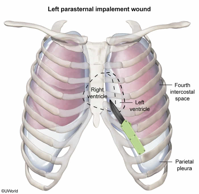
  • Right Ventricle
    • Site: Left sternal border (3rd–5th ICS).
    • Note: Most anterior chamber; #1 injured in cardiac stab wounds.


Penetrating Extremity Trauma

  • Epidemiology & Risk Factors
    • Gunshot wounds (GSW), stab wounds (knives, glass, shrapnel), industrial accidents.
    • Risk of vascular, neurologic, or osteoarticular compromise depends heavily on the mechanism/trajectory.
  • Clinical Features
    • Hard Signs of Vascular Injury:
      • Pulsatile bleeding.
      • Absent distal pulses.
      • Rapidly expanding hematoma.
      • Palpable thrill or audible bruit.
      • Signs of distal ischemia (6 Ps: Pain, Pallor, Pulselessness, Paresthesia, Paralysis, Poikilothermia).
    • Soft Signs of Vascular Injury:
      • Hx of massive hemorrhage at the scene.
      • Diminished (but palpable) distal pulses / asymmetric pulses.
      • Small, stable, non-expanding hematoma.
      • Neurologic deficit (adjacent nerve injury).
      • Proximity of injury to major vascular structures.
  • Diagnosis
    • Initial/Screening:
      • Hard signs present: Skip imaging -> OR immediately.
      • Soft signs present or asymptomatic: Ankle-Brachial Index (ABI) or Arterial Pressure Index (API) for upper extremity.
    • Key Imaging: Plain radiography (X-ray) of the extremity to assess for concurrent fractures, retained projectiles/foreign bodies, or subfascial air.
    • Confirmatory/Gold Standard: CT Angiography (CTA).
      • Indicated if ABI/API < 0.9, abnormal distal pulses, or soft signs present.
      • Duplex US is an alternative if CTA is unavailable or contraindicated.
  • Differential Diagnostics
    • Compartment Syndrome: Diff by tense, “wood-like” extremity, pain out of proportion, and severe pain with passive muscle stretch. (May co-exist or develop after reperfusion).
    • Isolated Peripheral Nerve Injury: Diff by focal motor/sensory deficits (e.g., foot drop) but normal vascular assessment (ABI > 0.9, normal pulses).
    • Isolated Orthopedic Trauma: Diff by bony deformity, crepitus, and positive X-ray findings without vascular compromise.
  • Management
    1. Stabilize (Primary Survey): ABCs. Control active hemorrhage with direct pressure. If direct pressure fails -> apply a tourniquet proximal to the injury.
    2. Surgical (Hard Signs): Immediate surgical exploration in the OR. Do not delay for imaging.
    3. Algorithmic Approach (Soft Signs / Asymptomatic):
      • Measure ABI/API.
      • If ABI/API < 0.9 -> CTA. (If CTA shows injury -> Surgery or endovascular repair).
      • If ABI/API ≥ 0.9 -> Serial physical exams + observation.
    4. Adjunctive Care:
      • Update Tetanus prophylaxis.
      • IV Abx (e.g., Cefazolin) if there is an open fracture or gross contamination.
      • Fracture stabilization (splint/traction) if applicable.
  • Complications
    • Acute limb ischemia -> gangrene -> amputation.
    • Compartment syndrome (often post-ischemic reperfusion; requires emergency fasciotomy).
    • Hemorrhagic shock / exsanguination.
    • Delayed vascular complications: Arteriovenous (AV) fistula, pseudoaneurysm.
    • Ischemic contracture (e.g., Volkmann ischemic contracture).
    • Osteomyelitis (if associated with open fracture or retained foreign body).

Penetrating abdominopelvic trauma

  • Immediate Indications for Exploratory Laparotomy (Skip Advanced Imaging):
    • Hemodynamic instability (HDI) (SBP < 90 mmHg).
    • Peritonitis (guarding, board-like rigidity, rebound tenderness). c
    • Evisceration (cover with sterile, saline-moistened gauze; do not reduce).
    • GSW with transperitoneal trajectory.
    • Impaled foreign object or active GI hemorrhage (blood per rectum, hematemesis).
  • Diagnostic Workup (Hemodynamically Stable Patients):
    • Anterior Abdominal Stab Wound:
      • Initial: Local Wound Exploration (LWE) in trauma bay.
      • Intact anterior rectus fascia Wound closure, tetanus update, discharge.
      • Violated or equivocal fascia Abdominal CT with IV contrast OR serial abdominal exams (q2–4h for 24–48h).
    • Flank / Back Wounds: Triple-contrast CT (IV, oral, rectal) to detect retroperitoneal injuries (duodenum, colon, kidneys).
    • Thoracoabdominal Wounds (Nipple to costal margin / 4th–8th ICS): Diagnostic laparoscopy (gold standard for occult diaphragmatic rupture).
    • Pelvic Penetrating Wounds: Contrast CT pelvis + CT cystography + rigid proctosigmoidoscopy (if blood on DRE).
    • eFAST Role: Quick bedside check for hemoperitoneum/pneumothorax, but negative FAST does NOT rule out hollow viscus, diaphragmatic, or retroperitoneal injury.
  • Acute Resuscitation & Medical Management:
    • Permissive Hypotension: Target SBP 80–90 mmHg (MAP ~65 mmHg) until surgical control (avoid if severe TBI).
    • Massive Transfusion Protocol (MTP): Early activation with a 1:1:1 ratio of PRBCs : FFP : Platelets.
    • Tranexamic Acid (TXA): Administer IV within 3 hours of injury.
    • Prophylaxis: Broad-spectrum IV antibiotics (e.g., Cefazolin + Metronidazole or Ampicillin-Sulbactam) + Tetanus toxoid TIG.
  • Surgical Strategy & Damage Control Surgery (DCS):
    • Triggered by the Lethal Triad: Hypothermia (< 35°C), Acidosis (pH < 7.2), and Coagulopathy.
    • Stage 1: Abbreviated laparotomy (stop hemorrhage, staple hollow-viscus leaks, pack 4 quadrants, leave abdomen open with negative-pressure dressing).
    • Stage 2: ICU resuscitation (rewarming, MTP correction of coagulopathy).
    • Stage 3: Definitive reconstruction and fascial closure in 24–48 hours.
  • High-Yield Complications & Distinctions:
    • Abdominal Compartment Syndrome (ACS):
      • Triad of bladder pressure > 20 mmHg + peak airway pressures + oliguria + hypotension/ CO.
      • Treatment: Immediate decompressive laparotomy.
    • Bladder Trauma Differentiation:
      • Intraperitoneal (Dome) Rupture: Peritonitis, urine ascites Immediate Ex-Lap.
      • Extraperitoneal (Trigone/Neck) Rupture: Associated with pelvic fractures Foley catheter drainage.