“Cherry-red” skin and mucous membranes (classic NBME buzzword; rare in vivo, typically a post-mortem finding).
Retinal hemorrhages (flame-shaped) on fundoscopy.
Tachypnea, tachycardia.
Diagnosis
Initial / Screening Trap:
Standard pulse oximetry (SpO2) is FALSELY NORMAL (conventional pulse oximeters cannot differentiate between oxyhemoglobin and COHb).
Confirmatory / Gold Standard:
Co-oximetry on ABG or VBG: Directly quantifies Carboxyhemoglobin (COHb) level.
COHb > 3% in non-smokers = Diagnostic.
COHb > 10–15% in chronic smokers = Diagnostic.
Key Laboratory Workup:
ABG: Normal PaO2, normal calculated SaO2, but severely reduced actual O2 content; anion gap metabolic acidosis (due to elevated lactic acid).
Cardiac Enzymes & ECG: Serial troponins and 12-lead ECG to rule out myocardial ischemia/infarction (CO is highly toxic to myocardium).
Serum Lactate & CK: Elevated lactate (tissue hypoxia); elevated CK (rhabdomyolysis in prolonged immobilization/coma).
Toxicology Screen & Pregnancy Test: Rule out co-ingestions; urine β-hCG in all females of childbearing age (fetus is at extremely high risk).
Imaging:
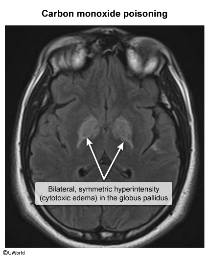
Brain CT/MRI: Bilateral, symmetric globus pallidus T2/FLAIR hyperintensity or hypodensity (characteristic ischemic necrosis). c
CXR: Normal or non-cardiogenic pulmonary edema in severe smoke exposure.
Differential Diagnostics
Cyanide Poisoning:
Diff: Also presents post-smoke inhalation with severe lactic acidosis (lactate>8–10 mmol/L), but COHb is normal (unless co-exposed). Breath smells like bitter almonds. Treated with hydroxocobalamin.
Methemoglobinemia:
Diff: Induced by oxidants (dapsone, benzocaine/lidocaine, nitrates). Cyanosis unresponsive to O2, “chocolate-brown blood”, SpO2 fixed around 85%. Diagnosis confirmed by elevated MetHb on co-oximetry. Treated with methylene blue.
Viral Gastroenteritis / Influenza:
Diff: Normal COHb levels. Symptoms persist outside the home and do not resolve with ambient fresh air.
Acute Coronary Syndrome (ACS):
Diff: May be triggered by CO toxicity in underlying CAD, but isolated ACS lacks co-oximetry elevation of COHb and basal ganglia involvement.