Epidemiology
Etiology
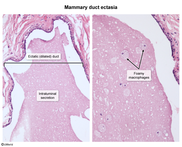
- A benign (non-cancerous) condition where large milk ducts beneath the nipple widen (ectasia), thicken, and fill with fluid.
- The exact cause is often unknown but is considered part of the normal aging process of the breast.
- Secretions can clog the duct, leading to periductal inflammation, fibrosis, and sometimes secondary infection.
- Smoking is a significant risk factor.
Pathophysiology
Inspissated luminal secretion → stasis → periductal inflammation → fibrous obliteration
Clinical features
- Most commonly affects perimenopausal and postmenopausal women (ages 45-55), but can occur at other ages.
- Often asymptomatic and found incidentally on mammogram.
- Classic symptom: Multicolored, sticky, thick nipple discharge (white, gray, green, or black) from one or both breasts.

- May present with a palpable subareolar lump, breast pain/tenderness, nipple retraction or inversion.
Diagnostics
- Ultrasound (US) is often the initial imaging modality to visualize dilated ducts. Ducts appear as dilated, branching tubular structures, sometimes with debris inside.
- Mammogram may show dilated ducts, benign-appearing calcifications (rod-like), or a subareolar mass.
- Biopsy may be needed if there is a suspicious mass or worrisome calcifications to exclude malignancy.