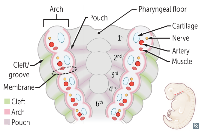
Pharyngeal clefts—derived from ectoderm. Also called pharyngeal grooves.
Just external ear canal
Pharyngeal arches—derived from mesoderm (muscles, arteries) and neural crest (bones, cartilage).
Connective tissue
Cartilage
Muscle
Bones
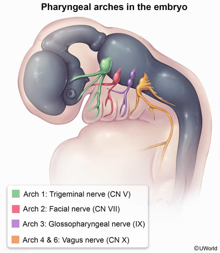
Nerves
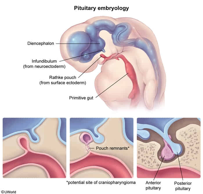
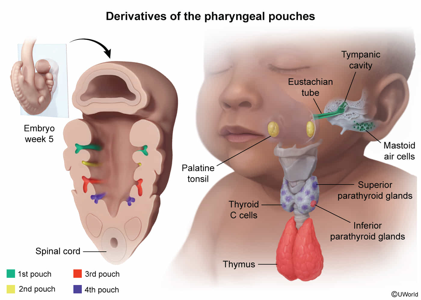
Pharyngeal pouches—derived from endoderm.
Epithelium
Glands
Mnemonic
CAP covers outside to inside:
Clefts = ectoderm
Arches = mesoderm + neural crest
Pouches = endoderm
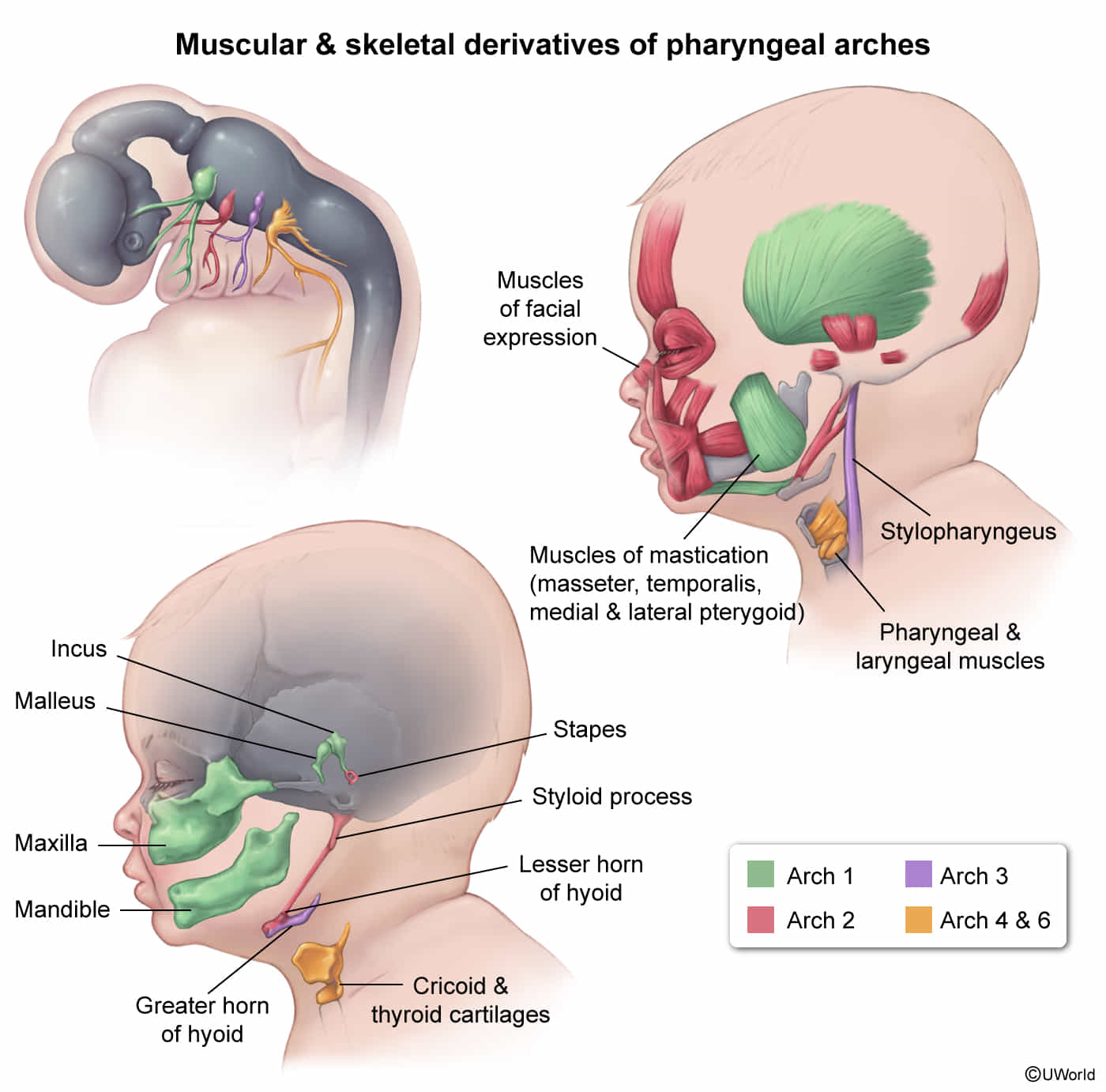
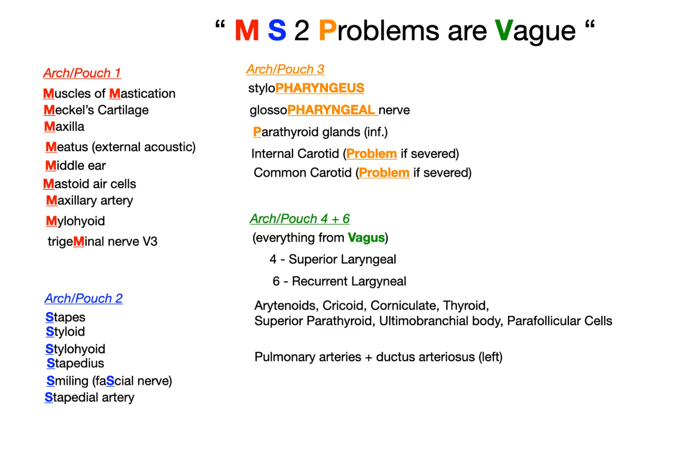
Pharyngeal arch derivatives
When at the restaurant of the golden arches, children tend to first chew (1), then smile (2), then swallow stylishly (3) or simply swallow (4), and then speak (6).
ARCH
NERVES
MUSCLES
CARTILAGE
1st pharyngeal arch
CN V₃ chew
Muscles of mastication (temporalis, masseter, lateral and medial pterygoids), mylohyoid, anterior belly of digastric, tensor tympani, anterior 2/3 of tongue, tensor veli palatini
Maxillary process → maxilla, zygomatic bone
Mandibular process → Meckel cartilage → mandible, malleus and incus, sphenomandibular ligament
 (Medical school 2 problems)