Ductal sex differentiation
- Mesonephric (Wolffian) duct → male internal structures
- Paramesonephric (Müllerian) duct → female internal structures
- Ureteric bud (from mesonephric duct) → ureter (in both genders)
Phases
1. Genetic & Gonadal Differentiation (Weeks 1-7)
- Indifferent Stage (until ~wk 7): Embryo has both mesonephric (Wolffian) and paramesonephric (Müllerian) ducts. Gonads are bipotential.
- Genetic Sex is determined at fertilization (XX or XY).
2. Male Development
- Key Driver: SRY gene on Y chromosome encodes for Testis-Determining Factor (TDF).
- TDF → Primitive gonads develop into testes.
- Testicular cells differentiate:
- Sertoli cells: Produce Müllerian Inhibitory Substance (MIS), also known as Anti-Müllerian Hormone (AMH).
- Function: Causes involution/degeneration of the paramesonephric (Müllerian) ducts.
- Absence of MIS → female internal structures develop.
- Leydig cells: Secrete testosterone.
- Function: Stimulates development of the mesonephric (Wolffian) ducts into male internal structures: Seminal vesicles, Epididymis, Ejaculatory duct, Ductus deferens (SEED).
- Sertoli cells: Produce Müllerian Inhibitory Substance (MIS), also known as Anti-Müllerian Hormone (AMH).
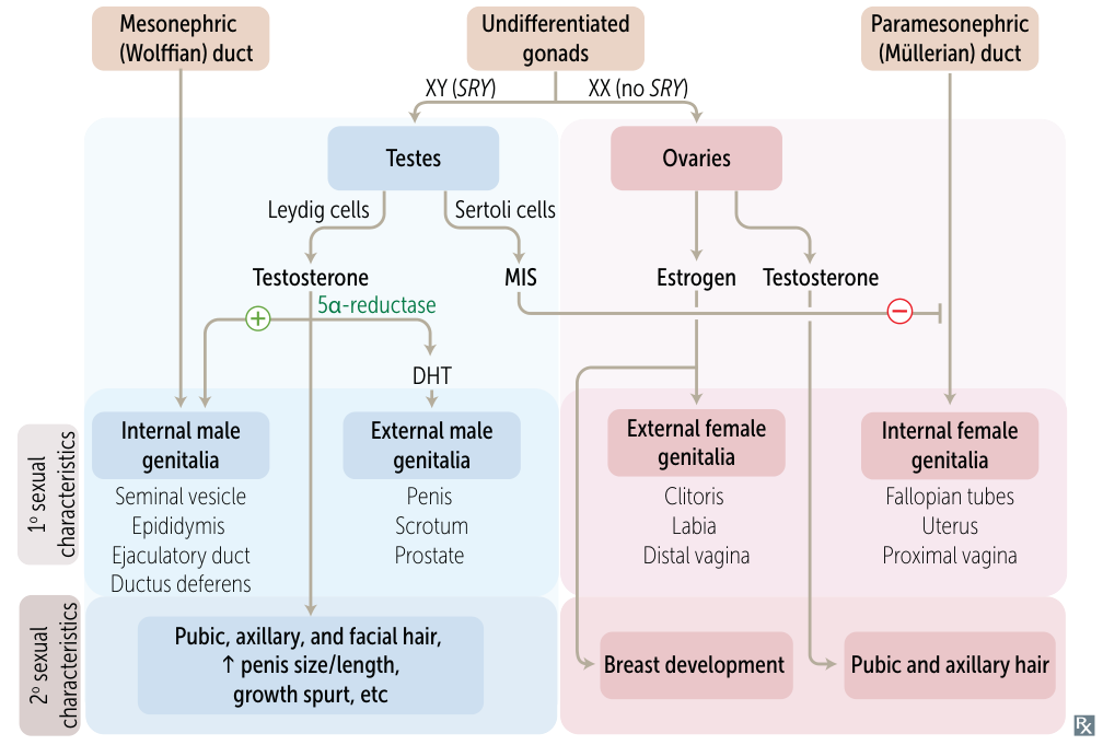
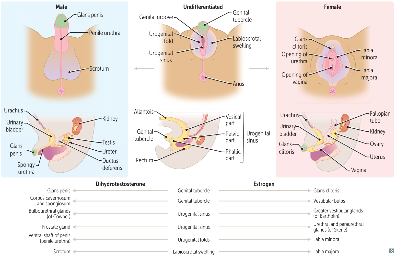
- External Genitalia: Testosterone is converted to dihydrotestosterone (DHT) by 5α-reductase. DHT is responsible for the development of male external genitalia (penis, scrotum) and prostate.
3. Female Development
- Key Driver: Absence of the SRY gene. Considered the “default” pathway.
- Gonads: Primitive gonads develop into ovaries.
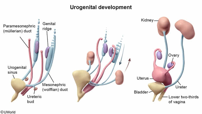
- Ductal System:
- No MIS/AMH from Sertoli cells → Paramesonephric (Müllerian) ducts persist and develop into female internal structures: Fallopian tubes, uterus, cervix, and upper 1/3 of the vagina.
- No testosterone from Leydig cells → Mesonephric (Wolffian) ducts degenerate.
- External Genitalia: Develop in the absence of DHT. The genital tubercle becomes the clitoris, urethral folds become the labia minora, and labioscrotal swellings become the labia majora.
4. Summary of Homologous Structures
Indifferent Structure | Male Derivative (stimulated by Androgens) | Female Derivative (default) |
---|---|---|
Gonad | Testis | Ovary |
Genital Tubercle | Glans penis, Corpus cavernosum/spongiosum | Glans clitoris, Vestibular bulbs |
Urogenital Sinus | Bladder, Urethra, Prostate, Bulbourethral glands | Bladder, Urethra, Lower 2/3 of vagina, Bartholin glands |
Urogenital Folds | Ventral shaft of penis (penile urethra) | Labia minora |
Labioscrotal Swellings | Scrotum | Labia majora |
Gubernaculum | Anchors testes to scrotum | Ovarian ligament, Round ligament of uterus |
External genitalia and urogenital differentiation
Uterine (Müllerian duct) anomalies
- General Impact
- ↓ fertility and ↑ risk of complicated pregnancy (eg, spontaneous abortion, prematurity, Fetal Growth Restriction (FGR), malpresentation).
- Diagnosis
- Hysterosalpingogram (HSG) can demonstrate uterine cavity shape and confirms tubal patency (via intraperitoneal spill of contrast).
- MRI is often the best imaging modality to differentiate specific anomalies.
Specific Anomalies
-
Septate uterus
- Patho: Incomplete resorption of the median septum after fusion of the Müllerian ducts.
- Notes: Most common Müllerian anomaly. Associated with the highest risk of miscarriage.
- Tx: Hysteroscopic septoplasty.
-
Bicornuate uterus
- Patho: Incomplete fusion of the Müllerian ducts.
- Notes: Results in a heart-shaped uterus with two horns.
-
Uterus didelphys
- Patho: Complete failure of Müllerian duct fusion.
- Notes: Results in a double uterus, double cervix, and often a double vagina.