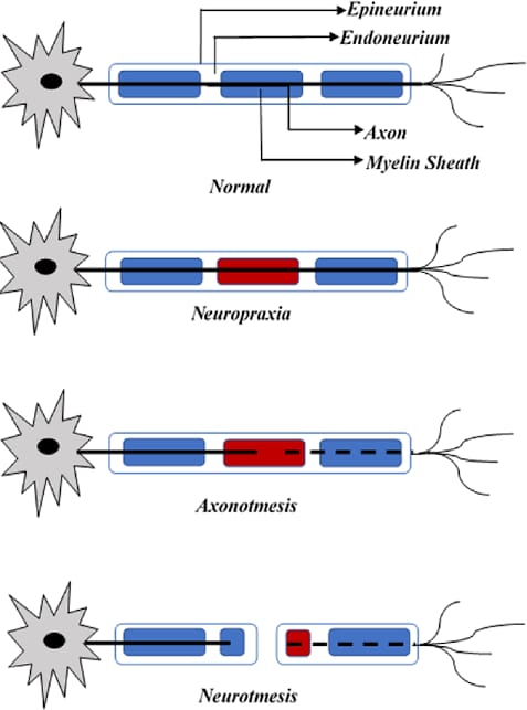
Types of Nerve Injury (Seddon’s Classification)
-
Neuropraxia (Mildest form)
- Endoneurium intact
- Axon intact
- Mild demyelination
- Causes mild conduction block leading to temporary weakness and sensory loss
- Complete, spontaneous recovery expected
- Examples: Crutch palsy, Saturday night palsy, stingers (sports injuries)
-
Axonotmesis (Moderate injury)
- Endoneurium intact
- Axon damaged/severed
- Moderate demyelination
- Moderate conduction block with more significant motor and sensory loss
- Good recovery possible without surgery
- Examples: Closed fractures, shoulder dislocations
-
Neurotmesis (Severe injury)
- Endoneurium damaged (possibly perineurium and epineurium too)
- Axon severed
- Severe demyelination
- Severe conduction block
- Poor recovery prognosis, typically requires surgical intervention
- Examples: Open fractures, deep lacerations, gunshot wounds
Nerve regeneration in axonotmesis
First degeneration, then regeneration!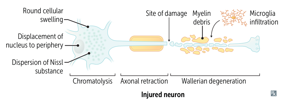
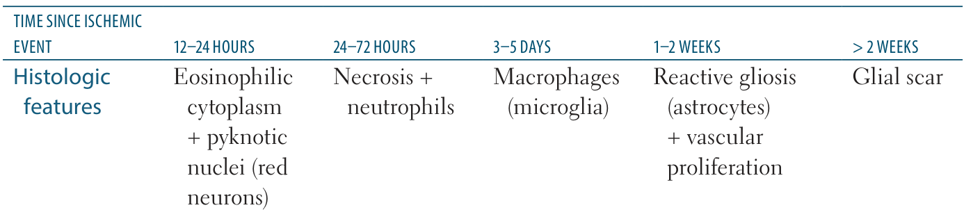
Only PNS nerve can regenerate, CNS nerves can only form glial scar.
Wallerian Degeneration Process
- Proximal Segment Changes:
- Chromatolysis: Nucleus shifts to periphery, Nissl bodies disperse, cell body swells
- Protein synthesis increases to support regeneration
- Breakdown of myelin and Schwann cells from injury site to adjacent upstream node of Ranvier
- Distal Segment Changes:
- Breakdown of axonal membrane, myelin sheaths, and some Schwann cells, from injury site to the nerve ending.
- Endoneurium releases chemicals (serotonin, histamine) attracting macrophages
- Macrophages clear axonal and myelin debris
- Schwann cells remain in the distal segment
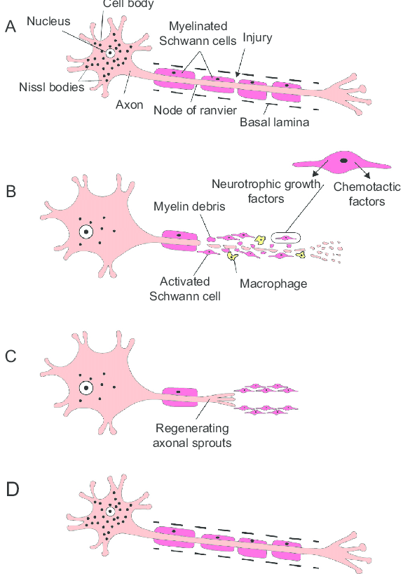
Nerve Regeneration Process
- Axonal sprouts form from proximal stump (within 24 hours)
- Sprouts grow toward distal stump at approximately 1.5mm per day
- Schwann cells cling to axonal sprouts and begin remyelination
- Proximal and distal stumps eventually reconnect
- Nucleus returns to center position in cell body
- Nissl bodies return to normal distribution
Leads to central chromatolysis
- Definition: the reaction of a neuronal cell body in response to an axonal injury
- Function: increase in protein synthesis to help restore the integrity of the damaged axon
- Characteristics
- Swelling of the neuronal body
- Dispersion of the Nissl bodies (which gives the name chromatolysis (chroma: color; lysis: disintegration))
- The rough endoplasmatic reticulum of neurons.
- Displacement of the nucleus to the periphery
Results in Wallerian degeneration
- Definition: an active neuronal degeneration process in response to axonal injury
- Function
- To clear axonal debris and prevent scarring
- Facilitate targeted reinnervation and functional recovery of tissues previously innervated by that axon before injury
- Characteristics
- Initially retained electrical excitability of axonal membrane distal to the injury, lasting up to 36 hours
- Progressive degeneration of distal segment cytoskeleton with dissolution of axonal membrane
- Degradation of residual myelin sheath by macrophages and Schwann cells
- In the central nervous system, phagocytic macrophages/microglia are recruited more slowly because of the blood-brain barrier. This slows removal of the myelin debris, which can persist for years in the degenerating tracts and suppress axonal growth via myelin-associated inhibitory factors.
- The proximal stump either stays in place or retracts slightly
- Ultimately, the cell body will sprout regenerative nerve fibers that, ideally, reinnervate the distal tissues.
- Regeneration is significantly more efficient in the peripheral nervous system than in the central nervous system.
- Good chance of at least partial recovery