Mnemonic
Think of muscle contraction as a climber (Myosin) trying to pull on a rope (Actin).
- Actin: The “A”ctive rope or track. It has binding sites (handholds) for myosin to grab.
- Myosin: The “M”otor protein. Its heads want to bind to actin and pull the rope to cause contraction.
However, two guards prevent the climber from grabbing the rope at rest:
- Tropomyosin: A long, “rope-like” protein that lies on top of the actin rope. It “traps” the myosin by physically blocking the binding sites. Think: Tropo-myo-sin = Stops Myosin.
- Troponin C: The “C”alcium-activated lock. It holds tropomyosin in its blocking position. It acts as the switch.
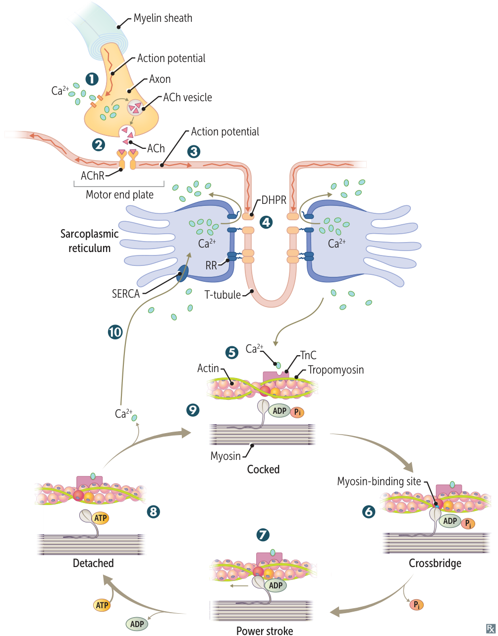
- Action potential opens presynaptic voltage-gated Ca2+ channels, inducing acetylcholine (ACh) release.
- Postsynaptic ACh binding leads to muscle cell depolarization at the motor end plate.
- Depolarization travels over the entire muscle cell and deep into the muscle via the T-tubules.
- Membrane depolarization induces conformational changes in the voltage-sensitive dihydropyridine receptor (DHPR) and its mechanically coupled ryanodine receptor (RR) → Ca2+ release from the sarcoplasmic reticulum (buffered by calsequestrin) into the cytoplasm.
- Tropomyosin is blocking myosin-binding sites on the actin filament. Released Ca2+ binds to troponin C (TnC), shifting tropomyosin to expose the myosin-binding sites.
- Myosin head binds strongly to actin (crossbridge). Pi released, initiating power stroke.
- During the power stroke, force is produced as myosin pulls on the thin filament A. Muscle shortening occurs, with shortening of H and I bands and between Z lines (HI, I’m wearing shortZ). The A band remains the same length (A band is Always the same length). ADP is released at the end of the power stroke.
- Binding of new ATP molecule causes detachment of myosin head from actin filament. Ca2+ is resequestered.
- ATP hydrolysis into ADP and Pi results in myosin head returning to high-energy position (cocked). The myosin head can bind to a new site on actin to form a crossbridge if Ca2+ remains available.
- Reuptake of calcium by sarco(endo)plasmic reticulum Ca2+ ATPase (SERCA) → muscle relaxation.
Sarcomere
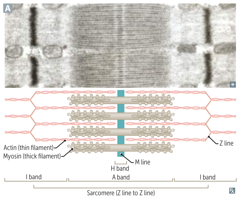
- Sarcomere: Functional unit, extends from Z line to Z line.
- Z line: A dark, zig-zag line that anchors the (+) ends of actin (thin) filaments. It forms the borders of the sarcomere.
- M line: A line in the Middle of the A band and H zone that anchors Myosin (thick) filaments.
- I band: Lighter band (I for Isotropic/light) containing only thin filaments (actin). It is bisected by the Z line.
- A band: Darker band (A for Anisotropic/dark) containing the entire length of the thick filaments (myosin), including regions of overlap with thin filaments.
- H zone: Lighter region in the middle of the A band (H for Helle, German for “bright”). It contains only thick filaments (myosin).