- Definition/Pathophysiology
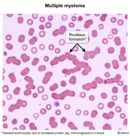
- Stacking of RBCs on a peripheral blood smear that resembles a “stack of coins”.
- Caused by high levels of circulating plasma proteins (e.g., monoclonal immunoglobulins, fibrinogen), which are positively charged.
- These proteins coat the negatively charged surface of RBCs, neutralizing the normal repulsive forces (zeta potential) between them and allowing them to aggregate.
Tip
For the USMLE, associate Rouleaux formation with the massive monoclonal protein spike of Multiple Myeloma. While SLE is a state of hypergammaglobulinemia, it is polyclonal and not of a high enough magnitude to be a characteristic finding.
- Key Associations (High-Yield)
- Multiple Myeloma (MM): This is the classic and most tested association. It is caused by the excessive production of monoclonal immunoglobulins (paraproteins or M-proteins) by malignant plasma cells.
- Waldenström’s Macroglobulinemia: Characterized by high levels of monoclonal IgM.
- Chronic Inflammatory States: Any condition that increases acute-phase reactants, particularly fibrinogen. This includes infections, malignancies, and connective tissue disorders. t
- Pregnancy: A physiologic increase in fibrinogen can lead to mild rouleaux formation.
- Lab Findings
- Markedly elevated Erythrocyte Sedimentation Rate (ESR) is a hallmark finding. The increased protein concentration causes RBCs to aggregate and settle faster.
- The background of the Wright-Giemsa stained smear may appear bluish due to high serum protein levels.
- DDx (on Peripheral Smear)
- RBC Agglutination: This involves irregular, 3-D grape-like clumping of RBCs, not neat stacks.
- Agglutination is typically caused by IgM antibodies cross-linking RBCs, as seen in cold agglutinin disease (e.g., secondary to Mycoplasma pneumoniae or infectious mononucleosis).
- Differentiating Test: Adding a drop of saline to the slide will disperse rouleaux, but true agglutination will persist.
- Clinical Workup
- The presence of rouleaux formation warrants an investigation for an underlying plasma cell disorder or other cause of hyperglobulinemia.
- Initial workup includes:
- Serum Protein Electrophoresis (SPEP) to identify a monoclonal (M-spike) or polyclonal gammopathy.
- Urine Protein Electrophoresis (UPEP) for Bence-Jones proteins.
- Serum free light chain assay.