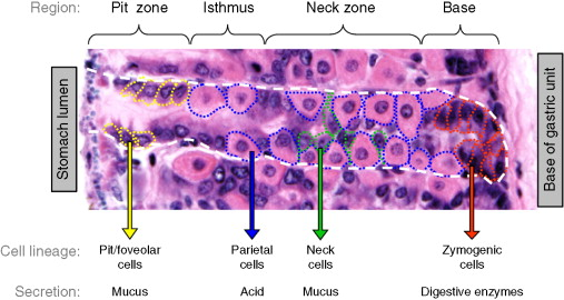
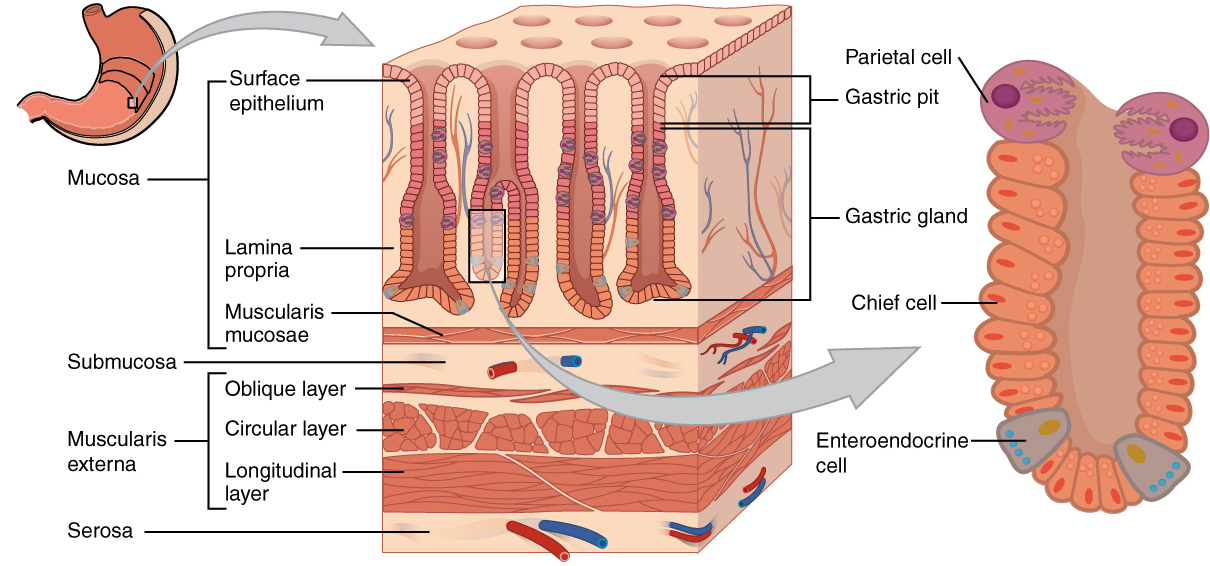
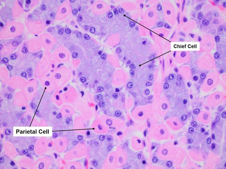
- Parietal Cells: Pink
- Location: Superficial/upper half of gastric glands (fundus and body).
- Appearance (H&E): Brightly eosinophilic (pink) due to abundant mitochondria required for H+ pumping.
- Size/Shape: Large, pyramidal or round/oval shape. Can have a “fried egg” appearance.
- Nucleus: Centrally located, spherical.
- Function: Secrete HCl (acid) and Intrinsic Factor.
- Chief Cells: Cyanosis (blue)
- Location: Deeper/basal half of gastric glands (fundus and body).
- Appearance (H&E): Densely basophilic (dark blue/purple) due to abundant rough endoplasmic reticulum (RER) for protein (pepsinogen) synthesis. Have pale, apical cytoplasm due to zymogen granules.
- Size/Shape: Smaller, cuboidal shape. Often found in clusters.
- Nucleus: Pushed towards the base of the cell.
- Function: Secrete pepsinogen.
GI hormones
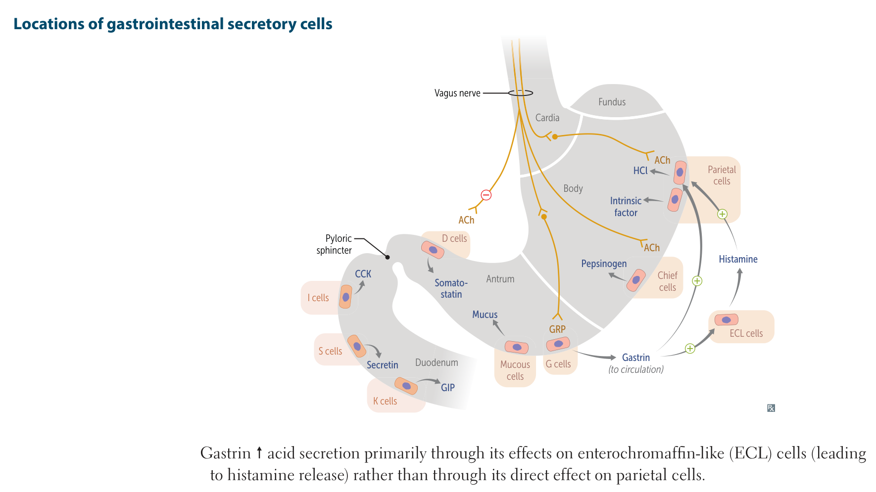
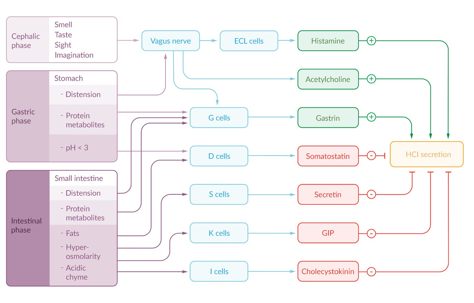
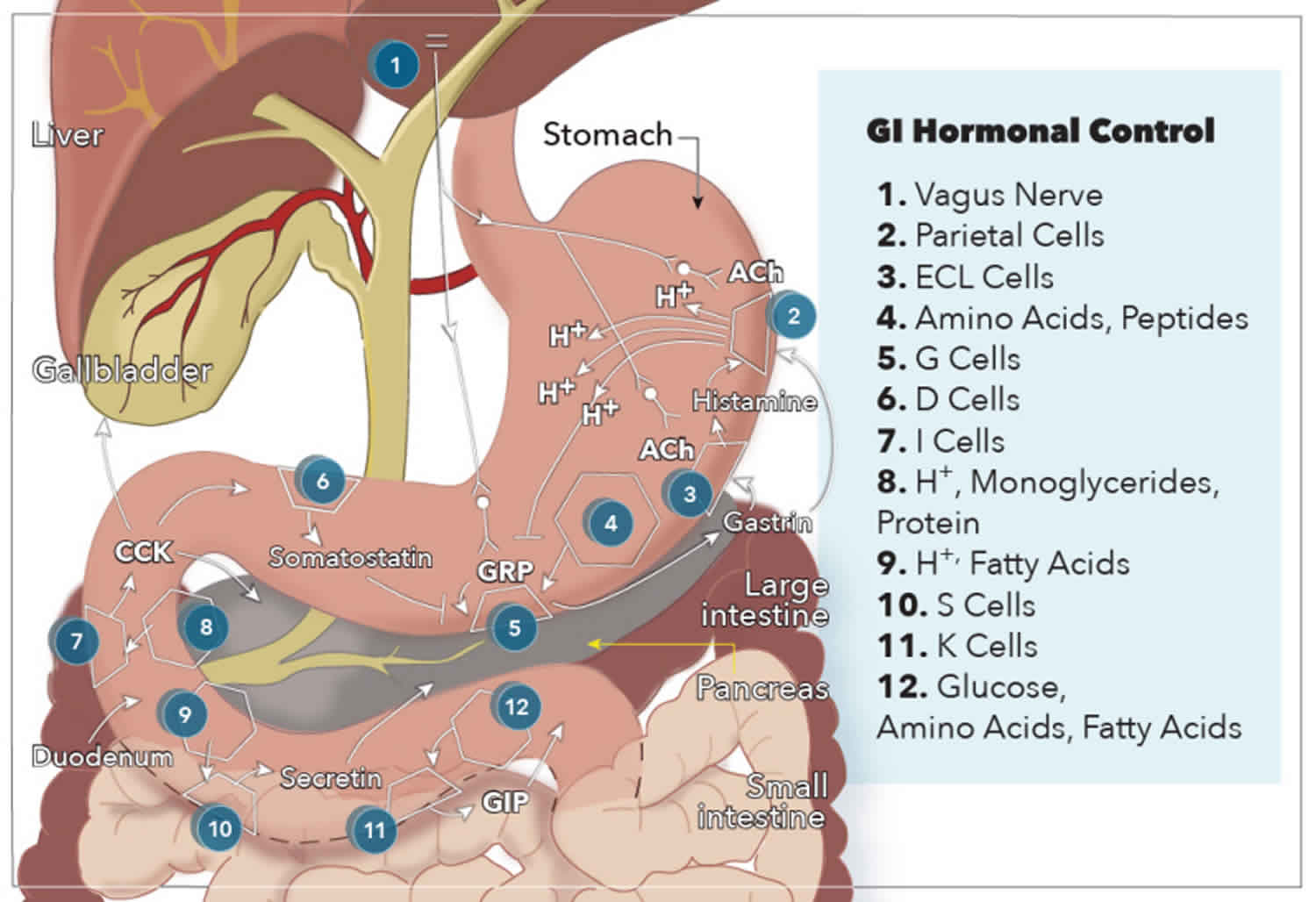
Mnemonic
- D cell → Somatostatin : “Death Star” kills all other hormones
- K cells “call to the Kitchen” GIP tells body dinner is ready
- I cells “Iron chef cells” CCK cuts up the molecules for digestion
- S cells “Stop right there” Secretin stops stomach acid production so the duodenum doesn’t get destroyed
Glucose-dependent Insulinotropic Peptide (GIP)
- Source: K cells (Duodenum, Jejunum).
- Triggers: Fatty acids, Amino acids, Oral Glucose. t
- Functions:
- Endocrine: Insulin release (Glucose-dependent).
- Exocrine: Gastric H secretion & motility.
- USMLE Buzzword: “Incretin Effect” t
- Oral glucose causes significantly higher insulin release than IV glucose because oral loads stimulate GIP/GLP-1.
- Clinical:
- Degraded by DPP-4.
- Tirzepatide: Dual GIP/GLP-1 agonist (Type 2 DM).