Gross Anatomy & Ligaments
- Hepatoduodenal Ligament: Connects liver to duodenum.
- Contains the Portal Triad: Proper Hepatic Artery, Portal Vein, Common Bile Duct.
- Pringle Maneuver: Surgical compression of this ligament to control hemorrhage. If bleeding persists, injury is likely to the IVC or Hepatic Veins (outflow vessels).
- Falciform Ligament: Connects liver to anterior abdominal wall.
- Contains Ligamentum teres hepatis (Round ligament) → remnant of fetal Umbilical Vein.
- Gastrohepatic Ligament: Connects liver to lesser curvature of stomach.
- Contains Gastric arteries.
- Functional Lobes: Right and Left lobes divided by Cantlie line (IVC to gallbladder fossa).
Blood Supply
- Dual Blood Supply:
- Portal Vein: ~75% of blood flow. Nutrient-rich, O2-poor. Drains GI tract, spleen, pancreas.
- Hepatic Artery: ~25% of blood flow. O2-rich. Branch of Celiac Trunk.
- Venous Drainage:
- Hepatic Veins (Right, Middle, Left) → IVC.
- Budd-Chiari Syndrome: Thrombosis of hepatic veins → centrilobular congestion/necrosis (Post-hepatic portal HTN).
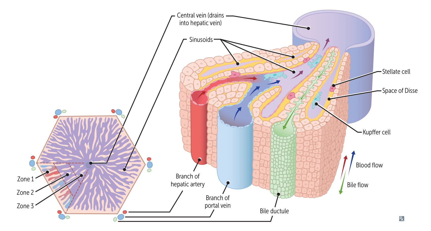
Liver zones
- Zone 1: The periportal zone
- Is best oxygenated and, therefore, is most resistant to ischemia.
- Affected first in viral hepatitis and toxic substance ingestion, e.g., cocaine.
- Zone 2: intermediate zone (liver): affected in yellow fever
Mnemonic
Zone II is affected in yellow fever.
- Zone 3: pericentral vein/centrilobular zone
- The least oxygenated zone, and thus most susceptible to ischemia
- Most sensitive to metabolic toxins (e.g., ethanol, CCl4, halothane, rifampin, acetaminophen)
- Site of alcoholic hepatitis
- Has the highest amount of cytochrome P-450
Tip
胆小管是肝细胞膜凹陷自然形成的,用来引流胆汁,所以只有增生没有缺如。