Epidemiology


Etiology

  • Cardiac cirrhosis
    • A complication of right-sided heart failure characterized by cirrhosis due to chronic hepatic vein congestion.
    • Associated with “nutmeg liver” (diffuse mottling on imaging due to ischemia and fatty degeneration).

Pathophysiology

  • Cytokine‑mediated activation of hepatic stellate cells has been identified as a central element for developing fibrosis.
    • A cell found in the perisinusoidal space of the liver
      • Quiescent phase: vitamin A storage
      • Activated phase: transform into myofibroblast to secrete collagen (primary cell involved in hepatic fibrosis)

Clinical features


Diagnostics

CT abdomen

  • Relative hypertrophy of the left lobe and caudate lobe
  • Regenerative nodules
  • Irregular liver surface
  • Indirect findings: ascites, splenomegaly, portosystemic collaterals

Pathology

  • Fibrosis (fibrous septa)
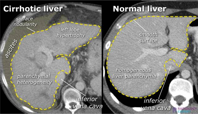
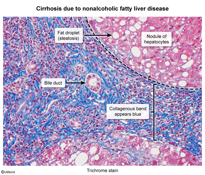
  • Replacement of normal liver tissue with collagenous regenerative nodules

Treatment


Complications


Portal hypertension

  • Portal-Caval Anastomoses
    • These are connections between the portal venous system and the systemic (caval) venous system. In portal hypertension, these sites become engorged and dilated as blood is shunted away from the high-pressure liver.
    1. Esophageal:
      • Portal: Left Gastric Vein
      • Caval: Esophageal branches of the Azygos Vein (drains to SVC)
      • Clinical: Esophageal Varices (risk of life-threatening hematemesis).
    2. Anorectal:
      • Portal: Superior Rectal Vein (from IMV)
      • Caval: Middle and Inferior Rectal Veins (drain to internal iliac vein → IVC)
      • Clinical: Anorectal Varices (often confused with hemorrhoids, can cause rectal bleeding).
    3. Paraumbilical:
      • Portal: Paraumbilical veins (travel with ligamentum teres)
      • Caval: Superficial Epigastric Veins of the anterior abdominal wall (drain to axillary/femoral veins → SVC/IVC)
      • Clinical: Caput Medusae (dilated, radiating periumbilical veins).
    4. Retroperitoneal:
      • Portal: Veins draining the ascending/descending colon, spleen, and liver.
      • Caval: Veins of the posterior abdominal/body wall (e.g., renal, lumbar veins).
      • Clinical: Usually asymptomatic, but can be a source of occult bleeding.
  • Management/Treatment
    • Treatment is aimed at managing and preventing life-threatening complications.
    • Primary Prophylaxis of Variceal Bleeding:
      • Non-selective β-blockers (e.g., propranolol, nadolol): Decrease portal pressure by reducing cardiac output and causing splanchnic vasoconstriction.
      • Endoscopic Variceal Ligation (EVL): Banding of varices to prevent rupture.
    • Acute Variceal Bleeding:
      • Medical Emergency: Secure airway, resuscitate with fluids/blood products.
      • Octreotide (somatostatin analog): Causes splanchnic vasoconstriction, reducing portal flow.
      • Urgent EGD: For banding/ligation of bleeding varices.
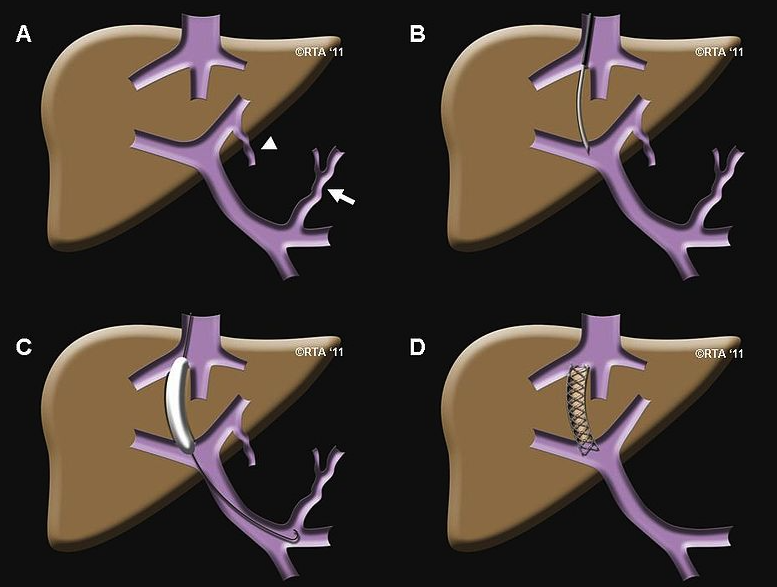
    • Refractory Bleeding/Ascites:
      • Transjugular Intrahepatic Portosystemic Shunt (TIPS): A stent placed between the portal vein and hepatic vein, shunting blood to decompress the portal system. Risk of worsening hepatic encephalopathy.
    • Ascites Management: Sodium restriction, diuretics (spironolactone, furosemide), and paracentesis for large volumes.
    • Definitive Treatment: Liver transplantation for end-stage liver disease.

Pulmonary complications of cirrhosis

Hepatic hydrothorax

  • Definition: pleural effusions (typically one-sided; 70% right, 18% left) with transudate characteristics in the absence of any other cardiac, pulmonary, or pleural disease
  • Pathophysiology: increased permeability of the diaphragm (small defects, increased abdominal pressure)
  • Clinical presentation
  • Diagnosis: Thoracocentesis shows transudate.