Epidemiology
Advanced age.
Female sex.
Obesity, sedentary lifestyle.
Prolonged standing/sitting.
Pregnancy.
Hx of DVT (Post-thrombotic syndrome ). c
Previous acute DVT causes inflammation and scarring of the delicate venous valves, leading to valvular incompetence, venous reflux, and sustained venous hypertension.
Fam hx of venous disease.
Pathophysiology : Incompetent venous valves → venous HTN → capillary leak of fluid/proteins/RBCs → tissue inflammation & fibrosis.
Etiology
Pathophysiology
Clinical features
Leg heaviness, aching, swelling (worse at end of day / prolonged standing; improves w/ elevation).
Varicose veins & telangiectasias.
Pitting edema.
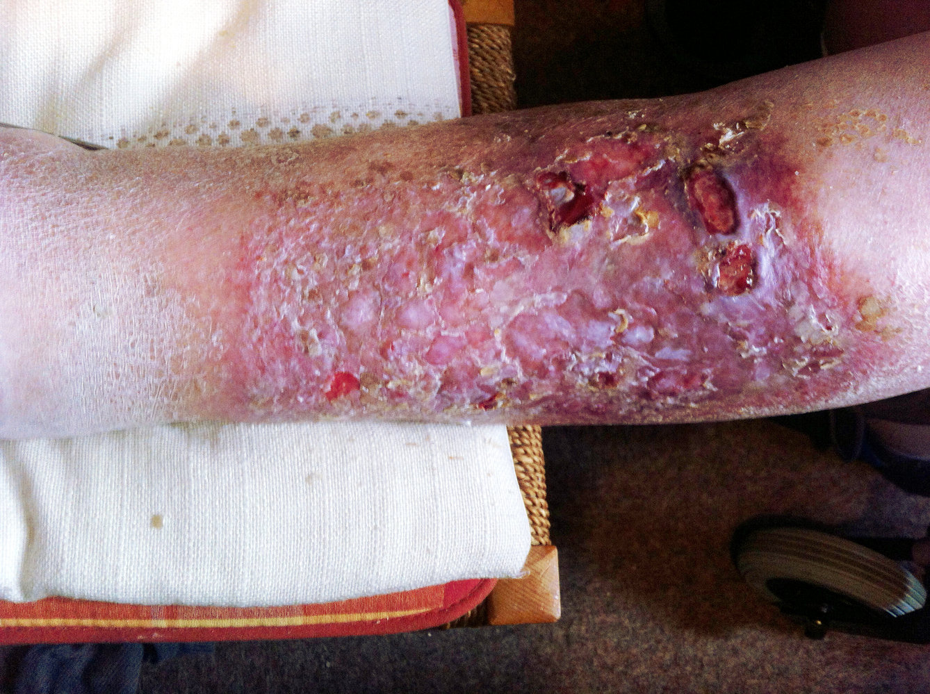
Stasis dermatitis : Erythema, scaling, pruritus of lower extremities.Hemosiderin deposition : Red-brown discoloration (from extravasated RBC breakdown).Lipodermatosclerosis : Subcutaneous fibrosis resulting in an “inverted champagne bottle” appearance of the lower leg.Venous stasis ulcers : Typically shallow, exudative, located at the medial malleolus .
Diagnostics
Initial : Clinical diagnosis based on typical H&P.Confirmatory/Best Test : Venous duplex ultrasound (evaluates for retrograde flow/venous reflux and rules out deep/superficial vein thrombosis).Note : Must evaluate arterial supply (ABI) prior to applying compression therapy.
Treatment
First-line (Conservative) :
Leg elevation (above heart level).
Graduated compression stockings (Contraindicated if concurrent severe PAD [ABI < 0.5]).
Weight loss, exercise (calf muscle pump).
Wound Care (for ulcers) :
Debridement of necrotic tissue.
Specialized dressings (e.g., Unna boot - zinc oxide-impregnated bandage).
Pentoxifylline or aspirin (may accelerate ulcer healing when added to compression).
Dermatitis Treatment : Mild-to-med potency topical corticosteroids for stasis dermatitis.Interventional/Refractory :
Endovenous thermal ablation (laser or radiofrequency).
Sclerotherapy (for telangiectasias/small varicosities).
Vein stripping/ligation (rarely used now).
Complications
Stasis dermatitis
Definition: eczematous dermatitis of the lower extremities caused by chronic venous hypertension and inflammation
Clinical features
Poorly defined erythematous, eczematous, and sometimes scaly patches commonly involving the medial malleolus Can involve pruritus; scratching may cause lichenification.
Acute stasis dermatitis can manifest with weeping, vesicles, and worsening erythema and edema.
Diagnostics
Perform diagnostic studies for CVI, including duplex US.
Consider skin biopsy if there is diagnostic uncertainty.
Extravasated erythrocytes, siderophages, and perivascular lymphocytes
Treatment
Initiate treatment of CVI, including compression therapy and referral for interventional treatment.
Identify and treat superimposed infections.