Cardiac auscultation
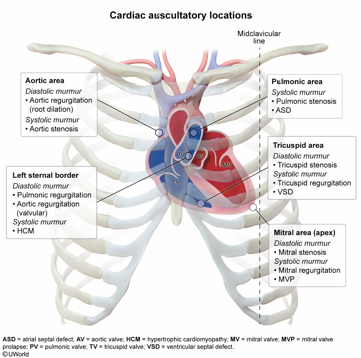

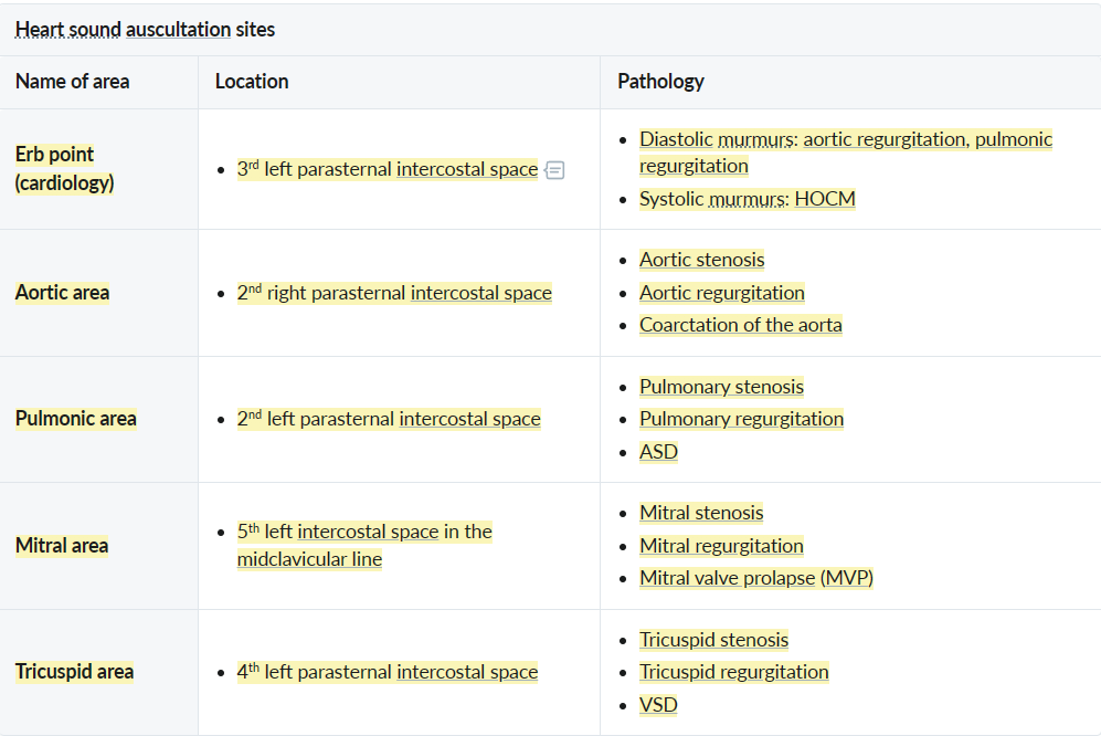
Heart murmurs

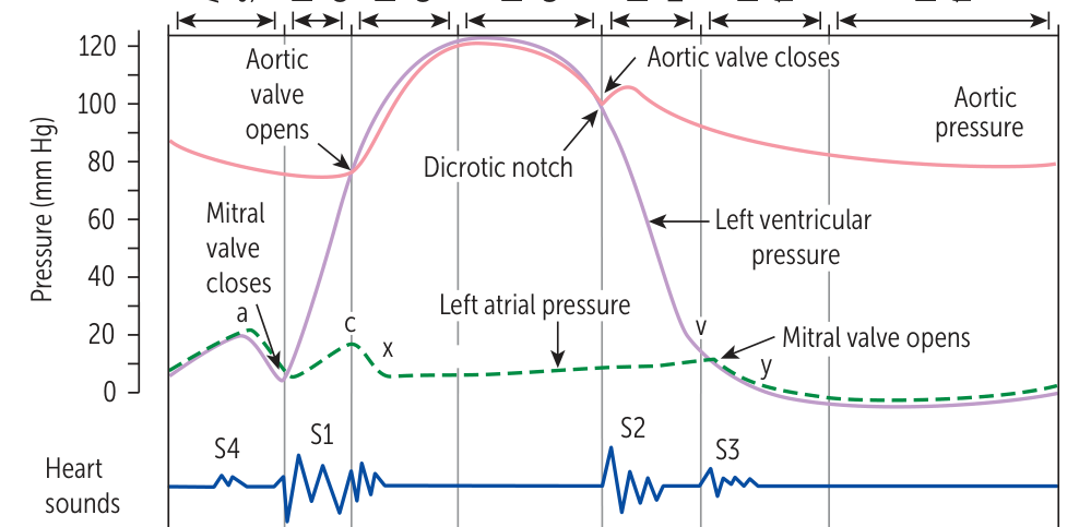
Heart sounds
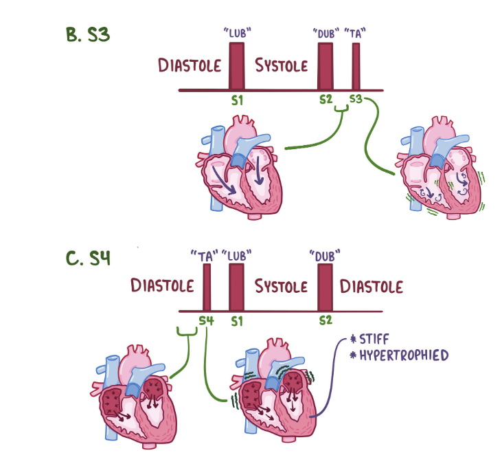
Extra (gallop) heart sounds
Tip
- S3: volume-overloaded
- Three tree → tree is big and large → ventricle is large
- S4: pressure-overloaded
- Four door → door is hard → ventricle is stiff
- Four door → door is hard → ventricle is stiff
S3
- Features
- Heard just after S2 (after opening of mitral valve)
- Caused by reverberant sound as blood fills an enlarged LV cavity during passive diastolic filling (i.e. end systolic volume is high)
- Associated disorders
- Heart failure with reduced EF
- High-output states (eg, thyrotoxicosis)
- Mitral or aortic regurgitation
- Teens or athletes
- Hearts are trained to handle more blood
S4
- Features
- Heard just before S1 (before closing of mitral valve)
- Caused by blood striking a stiff LV wall during atrial contraction
- As the atria contracts in late diastole against a stiffened ventricle, it must increase its force-production, which creates turbulent blood flow.
- Associated disorders
- Concentric LV hypertrophy
- Restrictive cardiomyopathy
- Acute myocardial infarction
S2 split
- Physiological split
- The sound of aortic valve closure (A2) precedes the sound of pulmonary valve closure (P2) during inspiration
- Especially pronounced among young individuals
- Wide split
- Caused by any condition that increases right ventricular afterload or decreases left ventricular preload
- Causes
- Pulmonary hypertension
- Pulmonary valve stenosis
- RBBB
- Fixed split
- ASD
- Paradoxical split (reversed split)
- Audible during expiration but not inspiration
- Expiration: A2 is heard after P2 during expiration due to delayed closure of the aortic valve (split reversal)
- Inspiration: the closure of the pulmonary valve is also delayed, resulting in A2 and P2 occurring simultaneously (i.e., a paradoxical decrease in the split during inspiration)
- Causes
- Aortic stenosis
- Left bundle branch block
- Audible during expiration but not inspiration
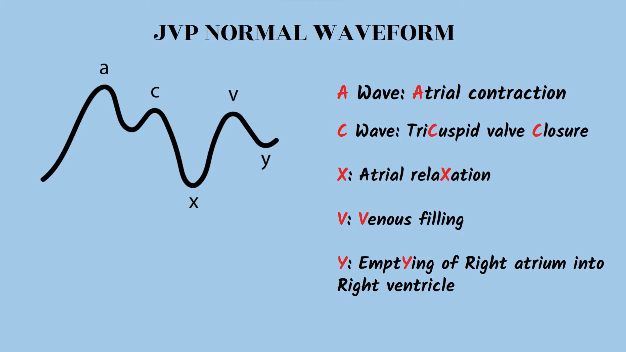
| Wave | Description | Abnormalities |
|---|---|---|
| a wave | The first peak caused by atrial contraction | Absent in atrial fibrillation |
| c wave | The second peak caused by tricuspid valve closure, contraction of the right ventricle, and bulging of the tricuspid valve into the right atrium | cv wave : severe tricuspid valve regurgitation |
| x descent | A drop in JVP caused by atrial relaxation | Absent in: |
| Tricuspid valve regurgitation | ||
| Right heart failure | ||
| v wave | The third peak caused by venous refilling of the right atrium against the closed tricuspid valve | Prominent in: |
| Tricuspid valve regurgitation | ||
| Right heart failure | ||
| y descent | A drop in JVP caused by decreased right atrial pressure as blood flows into the right ventricle after opening of the tricuspid valve | Prominent in: [7] |
| Tricuspid valve regurgitation | ||
| Constrictive pericarditis | ||
| Absent in: | ||
| Cardiac tamponade | ||
| Tricuspid valve stenosis |
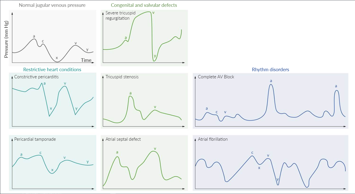
Pathology
Common abnormalities of the JVP waveform include:
- Constrictive pericarditis: elevated JVP (due to increased external atrial pressure) with a prominent x (exaggerated atrial relaxation) and y (early rapid ventricular filling) descent
- Cardiac tamponade: elevated JVP (due to increased external atrial pressure), a prominent x descent (exaggerated atrial relaxation), and a blunt or absent y descent (minimal ventricular filling)
- Tricuspid regurgitation: prominent v wave as the blood from the right ventricle regurgitates into the right atrium during ventricular systole (atrial diastole), increasing interatrial pressure and volume
- Tricuspid stenosis: giant a wave due to high right atrial systolic pressure
- Atrial septal defect: v wave ≥ a wave due to the left-to-right shunting of blood
- Third-degree atrioventricular (AV) block: cannon a waves due to the loss of AV synchronization and contraction of the atria against a closed tricuspid valve
- Atrial fibrillation: absent a waves due to ineffective contraction of the atria