Bernard-Soulier Syndrome
- Etiology/Pathophysiology
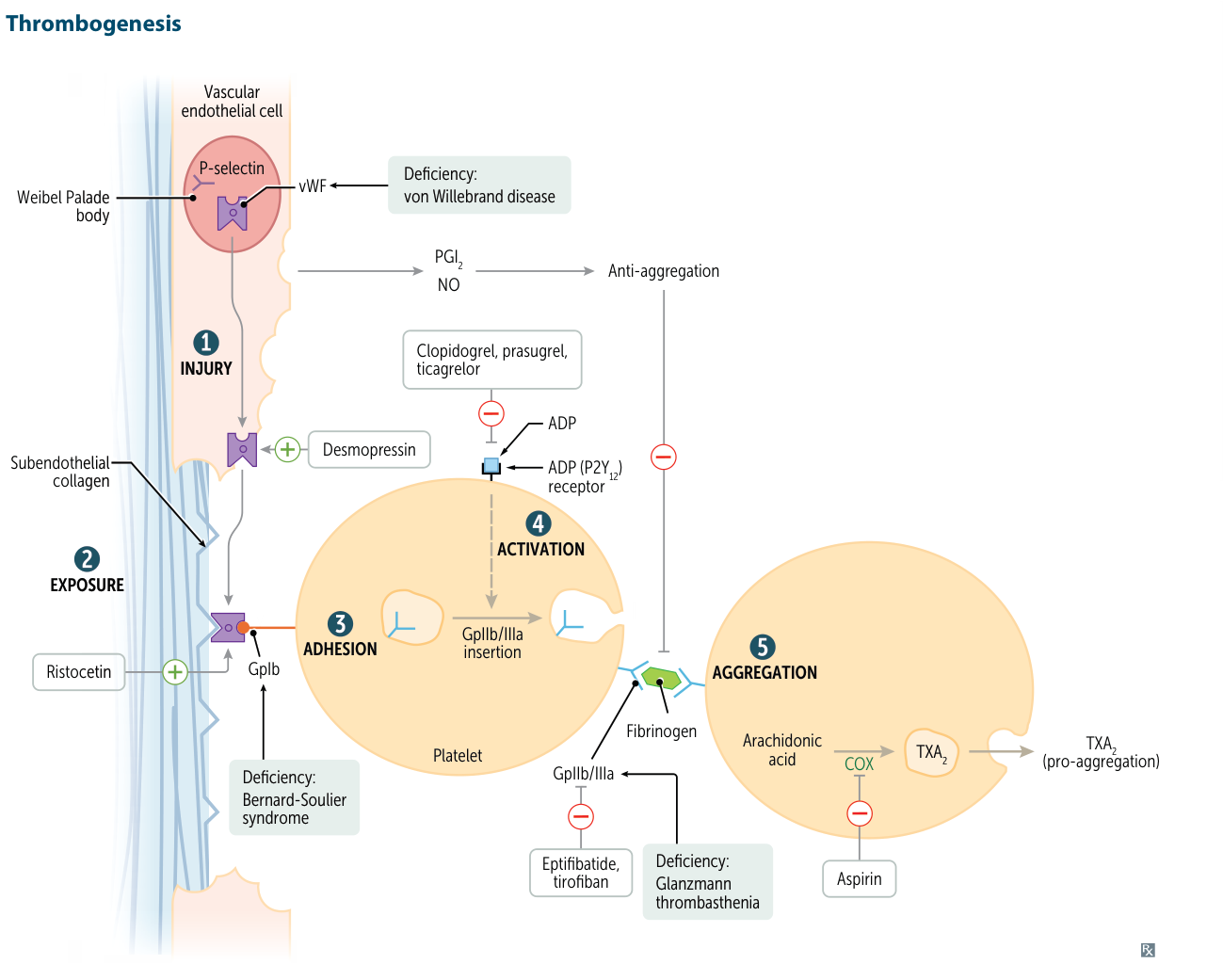
- Autosomal Recessive deficiency of GpIb.
- Impairs platelet Adhesion.
- Platelets cannot bind to vWF on exposed subendothelial collagen.
- Mnemonic: Bernard-Soulier = Big Suckers (Giant platelets) or Binds Surface (Adhesion).
- Clinical Features
- Mucocutaneous bleeding (epistaxis, menorrhagia, gingival bleeding).
- Petechiae, purpura, ecchymoses.
- Post-surgical bleeding/trauma.
- Diagnostics
- CBC: Mild to moderate Thrombocytopenia.
- Peripheral Smear: Giant Platelets (Large platelets).
- The GpIb-IX-V complex has two functions:
- External: Binds to vWF (Adhesion).
- Internal: Anchors the platelet membrane to the internal cytoskeleton (specifically Filamin A and actin).
- In Bernard-Soulier, the lack of GpIb disrupts the membrane-cytoskeleton link.
- The megakaryocyte cannot organize the cytoskeleton properly to pinch off small platelets.
- Result: Instead of small fragments, large, immature chunks of cytoplasm break off, resulting in Giant Platelets (often approaching the size of RBCs).
- Bleeding Time: ↑ (Prolonged).
- Ristocetin Cofactor Assay: Abnormal (No agglutination).
- Ristocetin activates vWF to bind GpIb; without GpIb, no clumping occurs.
- Does NOT correct with the addition of normal plasma (which contains functional vWF) (distinguishes from von Willebrand Disease). t
- Treatment
- Platelet transfusion (for severe bleeding).
- Antifibrinolytics (e.g., tranexamic acid).
Glanzmann Thrombasthenia
- Etiology/Pathophysiology
- Autosomal Recessive deficiency of GpIIb/IIIa.
- Impairs platelet Aggregation.
- Platelets cannot bind Fibrinogen (which links platelets together).
- Mnemonic: Glanzmann Aggregates (defect in aggregation).
- Clinical Features
- Identical mucocutaneous bleeding pattern to Bernard-Soulier.
- Petechiae, purpura, menorrhagia.
- Diagnostics
- CBC: Normal platelet count.
- Peripheral Smear: Normal platelet morphology; platelets appear isolated (no clumping).
- Bleeding Time: ↑ (Prolonged).
- Platelet Aggregation Studies:
- Abnormal: No aggregation with ADP, Epinephrine, or Collagen.
- Normal: Agglutination with Ristocetin (vWF-GpIb axis is intact).
- Treatment
- Platelet transfusion (severe bleeding).
- Recombinant Factor VIIa (refractory cases).
| Feature | von Willebrand (vWD) | Bernard-Soulier (BSS) | Glanzmann (GT) |
|---|
| Defect | vWF deficiency | GpIb deficiency | GpIIb/IIIa deficiency |
| Mechanism | Impaired Adhesion | Impaired Adhesion | Impaired Aggregation |
| Platelets | Normal count/size | ↓ Count, Giant size | Normal count/size |
| Ristocetin | Abnormal (Corrects w/ plasma) | Abnormal (No correction) | Normal |
| Key Diff | ↑ PTT (maybe) | Big Platelets (“Big Suckers”) | No clumping (Abnormal ADP agg) |